

What Brain Region Is Responsible For Learning Memory And Personality
What you'll larn to do: identify and describe the parts of the encephalon

In this section, you'll learn most the specific parts of the encephalon and their roles and functions. While this is non an anatomy class, you'll run into how important information technology is to understand the parts of the brain and what they practise and then that we can empathize mental processes and beliefs.
Watch IT
Watch this CrashCourse Psychology video for an overview on the brain and the interesting topics we'll embrace:
Learning Objectives
- Explain the ii hemispheres of the brain, lateralization and plasticity
- Place the location and function of the lobes of the brain
The Central Nervous System
The fundamental nervous organization (CNS), consists of the brain and the spinal cord.
The Brain
The encephalon is a remarkably circuitous organ comprised of billions of interconnected neurons and glia. It is a bilateral, or two-sided, structure that can be separated into distinct lobes. Each lobe is associated with sure types of functions, but, ultimately, all of the areas of the brain interact with one some other to provide the foundation for our thoughts and behaviors.
The Spinal Cord
It can be said that the spinal cord is what connects the encephalon to the outside world. Because of information technology, the brain tin act. The spinal cord is similar a relay station, just a very smart one. It not but routes messages to and from the encephalon, but it as well has its own system of automatic processes, called reflexes.
The top of the spinal cord merges with the encephalon stalk, where the basic processes of life are controlled, such as breathing and digestion. In the opposite direction, the spinal cord ends just below the ribs—contrary to what we might expect, it does not extend all the way to the base of the spine.
The spinal cord is functionally organized in 30 segments, corresponding with the vertebrae. Each segment is continued to a specific role of the body through the peripheral nervous system. Nerves co-operative out from the spine at each vertebra. Sensory nerves bring messages in; motor fretfulness send letters out to the muscles and organs. Messages travel to and from the encephalon through every segment.
Some sensory messages are immediately acted on by the spinal cord, without whatever input from the encephalon. Withdrawal from heat and knee jerk are two examples. When a sensory message meets certain parameters, the spinal string initiates an automated reflex. The betoken passes from the sensory nervus to a simple processing center, which initiates a motor command. Seconds are saved, because messages don't have to go the brain, be processed, and get sent dorsum. In matters of survival, the spinal reflexes allow the body to react extraordinarily fast.
The spinal cord is protected by bony vertebrae and cushioned in cerebrospinal fluid, only injuries nevertheless occur. When the spinal cord is damaged in a particular segment, all lower segments are cut off from the brain, causing paralysis. Therefore, the lower on the spine harm is, the fewer functions an injured individual loses.
The 2 Hemispheres
The surface of the brain, known as the cerebral cortex, is very uneven, characterized by a distinctive design of folds or bumps, known as gyri (singular: gyrus), and grooves, known as sulci (singular: sulcus), shown in Figure 1. These gyri and sulci form important landmarks that allow us to split the brain into functional centers. The about prominent sulcus, known every bit the longitudinal fissure, is the deep groove that separates the brain into two halves or hemispheres: the left hemisphere and the correct hemisphere.

Figure ane. The surface of the brain is covered with gyri and sulci. A deep sulcus is called a fissure, such as the longitudinal fissure that divides the brain into left and right hemispheres. (credit: modification of work past Bruce Blaus)
At that place is prove of some specialization of function—referred to as lateralization—in each hemisphere, mainly regarding differences in language power. Beyond that, still, the differences that take been found have been pocket-sized (this means that it is a myth that a person is either left-brained ascendant or right-brained dominant).[i] What nosotros exercise know is that the left hemisphere controls the right half of the body, and the right hemisphere controls the left one-half of the torso.
The two hemispheres are connected by a thick band of neural fibers known equally the corpus callosum, consisting of virtually 200 million axons. The corpus callosum allows the 2 hemispheres to communicate with each other and allows for information beingness processed on i side of the brain to be shared with the other side.
Normally, we are not aware of the different roles that our two hemispheres play in day-to-twenty-four hours functions, but there are people who come up to know the capabilities and functions of their two hemispheres quite well. In some cases of severe epilepsy, doctors elect to sever the corpus callosum equally a ways of decision-making the spread of seizures (Effigy two). While this is an effective treatment option, it results in individuals who have separate brains. After surgery, these split-brain patients bear witness a variety of interesting behaviors. For instance, a dissever-brain patient is unable to proper name a flick that is shown in the patient's left visual field because the information is only available in the largely nonverbal right hemisphere. However, they are able to recreate the picture with their left mitt, which is likewise controlled by the correct hemisphere. When the more verbal left hemisphere sees the picture that the hand drew, the patient is able to name information technology (assuming the left hemisphere can interpret what was drawn past the left hand).

Effigy two. (a, b) The corpus callosum connects the left and right hemispheres of the brain. (c) A scientist spreads this dissected sheep brain autonomously to bear witness the corpus callosum betwixt the hemispheres. (credit c: modification of work past Aaron Bornstein)
Much of what we know about the functions of dissimilar areas of the brain comes from studying changes in the behavior and ability of individuals who take suffered damage to the brain. For example, researchers study the behavioral changes caused by strokes to larn about the functions of specific brain areas. A stroke, caused past an suspension of claret flow to a region in the brain, causes a loss of brain function in the affected region. The harm can exist in a small-scale area, and, if it is, this gives researchers the opportunity to link whatsoever resulting behavioral changes to a specific area. The types of deficits displayed after a stroke will be largely dependent on where in the brain the damage occurred.
Consider Theona, an intelligent, self-sufficient adult female, who is 62 years erstwhile. Recently, she suffered a stroke in the front end portion of her right hemisphere. As a result, she has neat difficulty moving her left leg. (As you learned earlier, the right hemisphere controls the left side of the body; also, the brain'due south main motor centers are located at the front end of the head, in the frontal lobe.) Theona has also experienced behavioral changes. For example, while in the produce department of the grocery shop, she sometimes eats grapes, strawberries, and apples directly from their bins before paying for them. This behavior—which would accept been very embarrassing to her before the stroke—is consistent with harm in another region in the frontal lobe—the prefrontal cortex, which is associated with judgment, reasoning, and impulse control.
Link to Learning
Sentry this video to see an incredible example of the challenges facing a carve up-brain patient shortly post-obit the surgery to sever her corpus callosum.
Picket this second video about another patient who underwent a dramatic surgery to prevent her seizures. You'll learn more about the brain'due south power to modify, arrange, and reorganize itself, also known as brain plasticity.
Attempt It
Forebrain Structures

Figure three. The brain and its parts tin can be divided into three master categories: the forebrain, midbrain, and hindbrain.
Lobes of the Encephalon
The four lobes of the brain are the frontal, parietal, temporal, and occipital lobes (Effigy 4). The frontal lobe is located in the forward part of the brain, extending back to a scissure known as the central sulcus. The frontal lobe is involved in reasoning, motor control, emotion, and language. Information technology contains the motor cortex, which is involved in planning and coordinating movement; the prefrontal cortex, which is responsible for college-level cognitive functioning; and Broca's area, which is essential for language production.
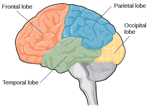
Figure 4. The lobes of the encephalon are shown.
People who suffer harm to Broca's area have great difficulty producing language of any form. For example, Padma was an electric engineer who was socially active and a caring, involved female parent. About twenty years ago, she was in a automobile accident and suffered harm to her Broca's area. She completely lost the power to speak and grade whatever kind of meaningful language. At that place is null wrong with her rima oris or her song cords, merely she is unable to produce words. She tin can follow directions but can't respond verbally, and she can read but no longer write. She tin can practice routine tasks like running to the market to buy milk, but she could not communicate verbally if a situation called for information technology.
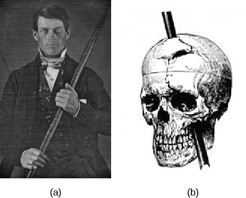
Figure 5. (a) Phineas Cuff holds the iron rod that penetrated his skull in an 1848 railroad structure accident. (b) Gage's prefrontal cortex was severely damaged in the left hemisphere. The rod entered Gage'southward face up on the left side, passed behind his eye, and exited through the top of his skull, before landing virtually 80 feet away. (credit a: modification of work by Jack and Beverly Wilgus)
Probably the nearly famous instance of frontal lobe damage is that of a man past the proper name of Phineas Gage. On September 13, 1848, Cuff (age 25) was working as a railroad foreman in Vermont. He and his coiffure were using an iron rod to tamp explosives down into a blasting pigsty to remove stone along the railway'southward path. Unfortunately, the iron rod created a spark and caused the rod to explode out of the blasting pigsty, into Gage'southward face, and through his skull (Figure 5). Although lying in a pool of his own blood with brain matter emerging from his head, Gage was conscious and able to get up, walk, and speak. Only in the months post-obit his blow, people noticed that his personality had changed. Many of his friends described him as no longer being himself. Before the accident, information technology was said that Cuff was a well-mannered, soft-spoken man, but he began to acquit in odd and inappropriate ways after the accident. Such changes in personality would exist consequent with loss of impulse control—a frontal lobe office.
Beyond the harm to the frontal lobe itself, subsequent investigations into the rod's path also identified probable damage to pathways between the frontal lobe and other brain structures, including the limbic organization. With connections between the planning functions of the frontal lobe and the emotional processes of the limbic arrangement severed, Gage had difficulty controlling his emotional impulses.
However, in that location is some prove suggesting that the dramatic changes in Gage's personality were exaggerated and embellished. Gage's case occurred in the midst of a 19th century debate over localization—regarding whether certain areas of the brain are associated with particular functions. On the basis of extremely express data near Gage, the extent of his injury, and his life earlier and after the accident, scientists tended to find support for their own views, on whichever side of the debate they fell (Macmillan, 1999).
Link to learning
Watch this prune about Phineas Gage to learn more than about his accident and injury.
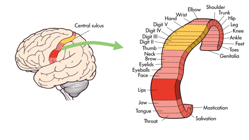
Figure 6. Specific trunk parts like the natural language or fingers are mapped onto certain areas of the brain including the chief motor cortex.
One specially fascinating surface area in the frontal lobe is chosen the "primary motor cortex". This strip running along the side of the encephalon is in charge of voluntary movements similar waving goodbye, wiggling your eyebrows, and kissing. It is an excellent example of the way that the various regions of the encephalon are highly specialized. Interestingly, each of our diverse trunk parts has a unique portion of the primary motor cortex devoted to it. Each private finger has about as much dedicated encephalon space as your entire leg. Your lips, in plough, require about as much dedicated encephalon processing as all of your fingers and your paw combined!
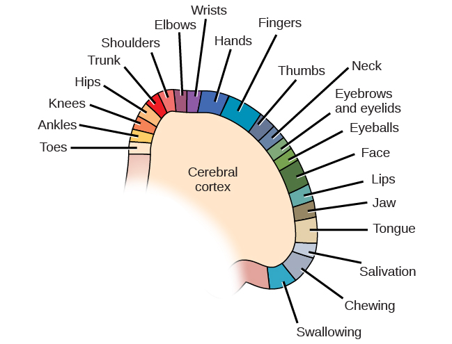
Figure 7. Spatial relationships in the body are mirrored in the organization of the somatosensory cortex.
Because the cognitive cortex in full general, and the frontal lobe in particular, are associated with such sophisticated functions as planning and being cocky-aware they are often thought of as a college, less primal portion of the brain. Indeed, other animals such as rats and kangaroos while they do have frontal regions of their brain exercise not have the aforementioned level of development in the cerebral cortices. The closer an animate being is to humans on the evolutionary tree—recollect chimpanzees and gorillas, the more developed is this portion of their encephalon.
The brain's parietal lobe is located immediately backside the frontal lobe, and is involved in processing information from the torso'south senses. It contains the somatosensory cortex, which is essential for processing sensory information from across the body, such equally touch, temperature, and pain. The somatosensory cortex is organized topographically, which means that spatial relationships that exist in the body are maintained on the surface of the somatosensory cortex. For instance, the portion of the cortex that processes sensory information from the hand is adjacent to the portion that processes information from the wrist.
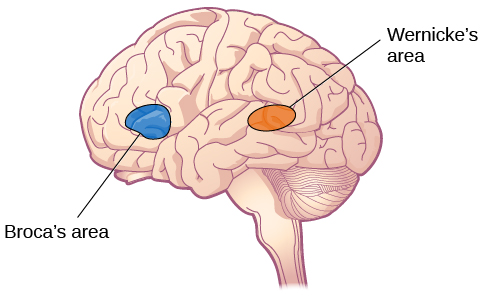
Figure viii. Damage to either Broca's area or Wernicke's area can event in language deficits. The types of deficits are very different, however, depending on which surface area is afflicted.
The temporal lobe is located on the side of the head (temporal ways "near the temples"), and is associated with hearing, memory, emotion, and some aspects of language. The auditory cortex, the main area responsible for processing auditory information, is located inside the temporal lobe. Wernicke's area, important for speech comprehension, is besides located here. Whereas individuals with damage to Broca'south area take difficulty producing language, those with harm to Wernicke'southward surface area tin produce sensible language, but they are unable to understand it.
The occipital lobe is located at the very back of the encephalon, and contains the primary visual cortex, which is responsible for interpreting incoming visual information. The occipital cortex is organized retinotopically, which means there is a shut relationship between the position of an object in a person'southward visual field and the position of that object'due south representation on the cortex. You will larn much more than about how visual information is candy in the occipital lobe when you report sensation and perception.
Try It
Food for Thought
Consider the following advice from Joseph LeDoux, a professor of neuroscience and psychology at New York University, every bit you learn about the specific parts of the brain:
Be suspicious of whatsoever statement that says a encephalon area is a heart responsible for some office. The notion of functions existence products of encephalon areas or centers is left over from the days when most show near brain function was based on the effects of brain lesions localized to specific areas. Today, we think of functions as products of systems rather than of areas. Neurons in areas contribute because they are part of a system. The amygdala, for example, contributes to threat detection because information technology is function of a threat detection system. And just because the amygdala contributes to threat detection does not hateful that threat detection is the only function to which information technology contributes. Amygdala neurons, for example, are likewise components of systems that process the significance of stimuli related to eating, drinking, sex, and addictive drugs.
Try Information technology
Glossary
auditory cortex:strip of cortex in the temporal lobe that is responsible for processing auditory information
Broca'south area:region in the left hemisphere that is essential for language production
cerebral cortex:surface of the brain that is associated with our highest mental capabilities
corpus callosum:thick band of neural fibers connecting the brain's two hemispheres
forebrain:largest part of the brain, containing the cognitive cortex, the thalamus, and the limbic system, among other structures
frontal lobe:part of the cerebral cortex involved in reasoning, motor control, emotion, and language; contains motor cortex
gyrus (plural: gyri): crash-land or ridge on the cognitive cortex
hemisphere:left or correct half of the brain
lateralization:concept that each hemisphere of the brain is associated with specialized functions
longitudinal fissure:deep groove in the brain'south cortex
motor cortex:strip of cortex involved in planning and coordinating move
occipital lobe:part of the cerebral cortex associated with visual processing; contains the primary visual cortex
parietal lobe:part of the cognitive cortex involved in processing various sensory and perceptual information; contains the primary somatosensory cortex
prefrontal cortex:expanse in the frontal lobe responsible for college-level cognitive functioning
somatosensory cortex:essential for processing sensory information from across the trunk, such every bit touch, temperature, and pain
sulcus(plural: sulci): depressions or grooves in the cerebral cortex
temporal lobe:part of cognitive cortex associated with hearing, memory, emotion, and some aspects of language; contains primary auditory cortex
Wernicke's area:important for speech comprehension

What Brain Region Is Responsible For Learning Memory And Personality,
Source: https://courses.lumenlearning.com/wmopen-psychology/chapter/outcome-parts-of-the-brain/
Posted by: robertsthenly.blogspot.com
0 Response to "What Brain Region Is Responsible For Learning Memory And Personality"
Post a Comment